The
Throwback Collection of Morgellons Specimens
(Read Jan's conclusions on top here, then her original article below, which laid the foundations for these conclusions. Make sure to keep up with her latest research by visiting her excellent website at: http://www.morgellonsexposed.com/ )
I wanted you to see the last part of the Throwback Collection that I have just revised. For the record
I will clearly state that transhumanization is the goal.No one component that is identified in Morgellons Disease is going to be a full answer. I have hesitated to talk about the transhumanism because some people are still stuck on even believing that we have a real disease. I guess it is time I made my position clear.
It took a long time to come to these conclusions and they are confirmed at every
turn by what I am finding. Jordan Maxwell had this all figured out a long time
ago. His video is just one of many things that confirm my beliefs beyond my own
findings:
https://www.youtube.com/watch?v=5jJKue2Ff6o
Last part of the "Throwback Collection"
Along with that we have the presence of Beta-1,3-Glucans from the oomycota varieties which are known for their anti-tumor effects and beneficial
effects on the immune system. How nice of them to throw out a health bonus. As I have just mentioned it is also known for it's ability to produce an environment for the
development of nano machines and bio-engineering. Take a look at my photo journal and see the many categories of wires and components inhabiting the human body. This is
not random.
Consider the implications of the bigger picture and the fact that most people without symptoms have had the same exposure to the same chemtrails
and tainted food as Morgellons victims. These people may simply be assimilating the technology without problems or symptoms. Morgellons disease may actually be the lack
of ability to assimilate the crap they are dumping on us due to an aberration in our immune systems or our genetics. This aberration was probably unforeseen by those who
are doing this to us. As such it is an unwelcome circumstance that is being ignored, ridiculed and constantly monitored by professionals. Reality is not always apparent
until it is in hindsight. Further, all "people" we meet are not necessarily what they appear to be.
This is not a one-component disease but a system of synergistic pathogens with a purpose.
As an update, I want the public to understand that there is no valid funded research being done by our government. A handful of people are
trying to make progress with their own time and money but it seems that there is a brick wall placed in front of us at every turn. I believe the term for this is that
we are being "contained". A number of medical professionals have found that involvement with the Morgellons issue has led to government shakedowns of their records in
attempts to find any reason to close them down. Other researchers have been threatened and more.
There is a dark underbelly of evil that is calling the shots regarding
this man-made disease. It is time to wake up and discern the truth.
Points to ponder:
.........Could it be that some symbiotic balance is being kept in the grand scheme of things to create a favorable environment for the dictyostelium,
oomycetes and other genetic bio-mixtures to live well and prosper? In the bigger picture most things found are serving a purpose and act together. These are not all
random things thrown into the mix without thought or purpose. To find the answers, all of the components must be looked at as a whole system.......
Original Article:
In the first three years I suffered with Morgellons disease I
was totally on my own. All I knew was that I had something on and in my
body that I could see and feel but my doctors were trying to tell me I was
having delusions of parasites. They also told me the ulcerous sores on my
body were the results of self excoriation. I made all of the classic
moves like bringing in fibers to show the them. The humiliating treatment
I received from the medical profession only succeeded in making me very angry
and fueled my great determination to prove them wrong. I thought that I
was the only person going through this misery and didn't learn for several
years that other had these same symptoms.
In the beginning I was looking for something with a
logical and biological explanation. I was not into any sort of
conspiracy theories. I simply thought I would get a microscope and study
what was on my body. I believed that with enough studying and
looking through the scope that I would identify what disease I had from a known
database of recognized diseases. I began a 3 year study of all fungus,
cyanobacteria, chytrids, rotifirs, algae or anything else I could learn about
that might be a clue to this mystery.
Eventually I looked at the Internet and typed some symptoms into
a search engine. I was amazed and excited that I found Mary Leitao's
Morgellons website. I contacted Mary and started working with her.
I asked her about the name �Morgellons�. She had taken it from a
1690 monograph by Sir Thomas Brown who described a condition in children that
included copious growth of hair on their backs. I can't say that I agreed
with her choice to use that particular name for this illness but I supposed it
sounded more medically correct than what I have been calling it which was
�the crap.�
During the time I worked with Mary she was able to get some of
my Morgellons samples into a lab at the University of Pittsburgh. They
underwent testing using Fourier Transform Infrared Spectroscopy. They
were found to be hollow, auto fluorescent and to contain cellulose but no
chitin. From my earlier study of mycology I knew that the absence chitin
ruled out any fungus since all fungus contains chitin. At that time I got
a letter from Mary and with some micrograph images taken by the lab. Due
to a computer assault from an evil anti-Morgellons website I no
longer have copies of these. Apparently the samples were quite a
sensation at the lab and could not be readily identified. There were to be
further studies done and frozen cross sections for further testing had already
prepared by the lab. Shortly thereafter I learned that the University had
told Mary they had no time to work on it. This was surprising since it
was a unique material with many unanswered questions and this was, after all, a
University where I would hope knowledge would be valued. I began to
wonder why the Morgellons research had been dumped like a hot
potato.
Several years later more fiber samples from my body were
determined to be made of HDPE ( high-density polyethylene) by another
researcher who paid to have them tested at several accredited labs. There
are differences in the fibers since some are round and some are more
ribbon-shaped. There may also be a difference if a fiber directly enters
the body from the environment or if it is a second or more generation product
of self replication that occurs in the body.
In later years it was apparent to me that the bizarre nature of
my Morgellons findings were unnatural and likely from a man-made source.
It was not a pleasant journey as I began to "wake up" to what
was happening in this altered reality. Much to my dismay, all roads
carried me to the same conclusion and still do. The pathogens in my
body came from a bio-lab and have the hand of man written all over them.
It occurred to me that if these pathogens were being bioengineered in a
lab that they were made of multiple components and that the mutated material
might reproduce and intermittently send out a batch of identifiable debris much
like the original genetics. I call this type of debris
�throwbacks�
(http://www.morgellonsexposed.com/Throwback.htm).
The multi-component part of my theory has
proven to be true.
See my paper entitled:
"Revelations From a Man Who Helped
Design Morgellons Disease"
(http://www.morgellonsexposed.com/RevelationsOfaMan.htm)
(RE: Dr Gwen Scott and her admissions from a
government scientist)
It is my theory that when new life forms or designer diseases
are created in bio-labs using the processes of gene splicing, mutation,
bioengineering or nanotechnology that components of these mixtures can self
replicate with varied results. Components such as dictyolstelium
discoidium, insect genes, and other pathogens will continue to make some new
mutations in each generation of self replication. I believe that some of
these mutations "throw back" to expressions similar to the original
materials used in the lab created mixture. Specimens from Morgellons
disease tissue can sometimes be identified as nearly identical to the original
components used in this biological witches brew before the mutation
process. It is not unlike finding a child with flaming red hair in a
family of people who do not have this characteristic. Hopefully the
previous ancestral hair colors of past generations were known to the parents
since situations like this can lead to being a guest on The Maury Povich (Who
my Baby Daddy?) DNA Testing Show. If only gene identification for
Morgellons disease was this easy.
The information in this paper is only my personal theory but it
is the best explanation I can find to make sense of all of the strange
artifacts that come from people with Morgellons disease. It is also why I
think that some of these strange findings occur most frequently in the early
phases of the disease and do not repeat as often in later years of the
disease. I will later discuss a strange cellular slime mold
known as dictyostelium discoidium. It is all about using the genetic
characteristics of this cellular slime mold to produce mutations in a bio-lab.
I believe it is one of many parts of the Morgellons story.
Most of my �throwback� findings only appeared in the beginning
of the disease when I had multiple lesions and things were at their worst.
That was about 7 years ago. I had tentatively identified some of
the debris as members of 3 varieties of the Oomycota phylum of pseudo fungus.
There are over 500 varieties and many of them can have ill effects of humans,
animals and plants.
The varieties of oomycota can produce diseases like pythiosis,
lagenidosis, rhinosporidosis, downy mildew, blue tobacco mold, the fish disease
saprolegnia (Ich) and the infamous phytophthora infestans that had killed
millions in the Irish Potato Famine by blighting the spuds.
I had also found bizarre mutations of earthworms and other
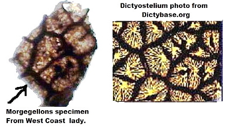
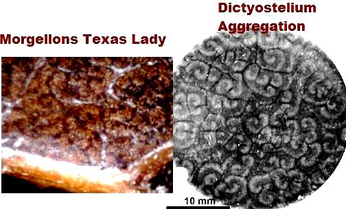
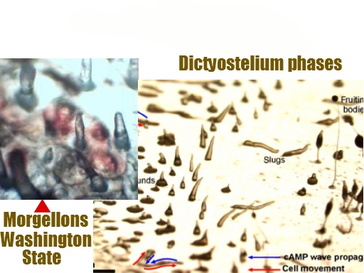
material that looked like dictyostelium discoidium. The reason that the
dictyostelium discoidium is important is because this cellular slime mold
has unique properties that lend itself to mutating other substances.
These eukaryotic microorganisms have a simplistic genetic makeup and produces
what are know as chemotaxis. Dictyostelium is utilized in many labs to
specifically mutate other material. Dictyostelium has an amoeba form
along with many other stages of varied shapes and configurations as the
individual cells emerge into groupings that look like larger single entities.
�Dictyostelium amoebae grow as separate, independent cells but
interact to form multicellular structures when challenged by adverse conditions
such as starvation. Up to 100,000 cells signal each other by releasing
the chemoattractant cAMP and aggregate together by chemotaxis to form a mound
that is surrounded by an extracellular matrix. Processes depend on
cell-cell communication in Dictyostelium. Many of the underlying
molecular and cellular processes appear to have arisen in primitive precursor
cells and to have remained fundamentally unchanged throughout evolution.
Basic processes of development such as differential cell sorting, pattern
formation, stimulus-induced gene expression, and cell-type regulation are
common to Dictyostelium. It is used in gene research as well as other
uses� (dictybase.org).
Even if you have never before heard of dictyostelium discoidium
you may be quite impressed to go to this huge website that is well funded and
part of the Human Genome Project and NIH. This cellular slime mold
is a major player in many aspects of medicine and cell research. It is a
good bit of information to have for future reference. I encourage
everyone to take a look at photos and videos of this substance at
http://dictybase.org/
To watch these little amoeba squirm and aggregate watch video
here:
http://dictybase.org/Multimedia/development/agg.mpg.
It may have an eerie familiarity to Morgellons victims.
The many
configurations of Dictyostelium:
Each shape is comprised of hundreds to thousands of single
motile amoeba cells acting in unison to form each of these configurations.
~~~~~~~~~~~~~~~~~~~~~~~~~~~~~~~~~~~~~~~~~~
From: "Texas
Medical Center News"
Vol. 21,
No. 18 October 1, 1999
Genome Studies
of Slug Might be Applicable to Humans
"Dictyostelium consists of only six chromosomes and approximately
8,000 genes. The DNA in those genes comprises 34 million pairs of
chemicals called bases that contain instructions for the role each gene plays.
"
development, Dr. Kuspa can
determine the gene's function. It takes only a month to generate a
mutation in Dictyostelium, but in a more complicated genome, such as that of a
mouse, the process can require six months to a year.
~~~~~~~~~~~~~~~~~~~~~~~~~~~~~~~~~~~~~~~~~~
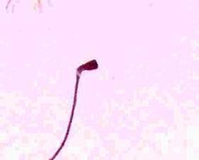
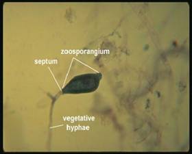
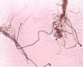
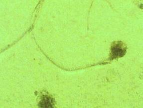
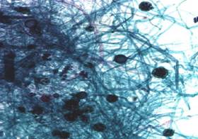
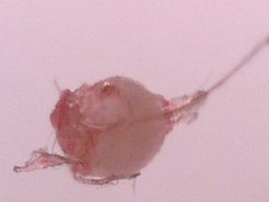
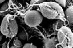
Here are some of the �throwbacks� I found in my own tissue from
the phylum oomycota (o-o-my-CO-ta) 3 photos of saprolegnia
zoosporangium.
|
1.Saprolegnia from my
body 200x |
Saprolegnia
oospores web photo
|
|
2. Saprolegnia zoosporangium 60x
|
3. Cluster saprolegnia 200x
|
These are not the original coloration as shown except for small
image on lower left fibers were dark blue, bulbs on the ends of fibers
were brown/red. The bulbs (zoosporangium) would fall off as a separate
component. I only found this formation twice, about 5 years ago.
|
Saprolegnia
oospores - 200x |
Saprolegnia oospores
web photo
|
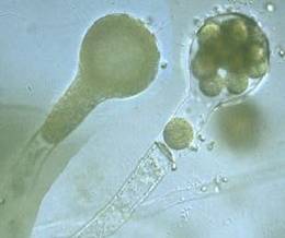
The photo below is not from my body but I did find a specimen
exactly like this. I was able to get a drop of the clear sticky liquid
that came out of a lesion onto a slide. At a magnification of 350x I
found a tiny but uniquely distinct shape on the slide. I could not then
identify it but I described it to Mary Leitao as a clear snake skin with tiny
round circles in it. I was sure this slide would be a breakthrough for the
research but unfortunately that did not happen.
I did not have a microscope that I could take a digital image
with at that time. I did have a scientific microscope. I sent this
slide to Mary Leitao who is a trained biologist. She looked at it and
acknowledged the specimen on the slide. She forwarded that slide and many
others to Dr. William Harvey who was then in private practice. The slides
were said to have been �lost� during a move by his office. Formerly Dr.
Harvey had worked for NASA full time and at that time was still the Chairman of
their Educational Advisory Board. I must admit that I still question what
happened to my slides. I later identified the missing specimen as
Dictyuchus from a web photo.
|
Dictyuchus Species of Oomycota
|
All of the varieties of oomycota I have mentioned thus far
are related to the fish disease known as Ich or Saprolegnia. I do not
believe that Morgellons victims have a pure strain of this disease but I do
believe that some of the genetics were used from this disease in the
bioengineered mixture.
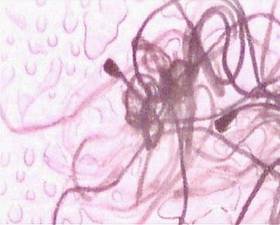
|
|
Saprolegnia Fibers in Nature (blue and red)
|
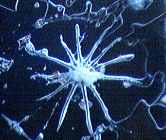
I believe that other varieties of the oomycota family have been
put into the mix as well. I have previously found this star fish shape in
my lesions. I know that others have found them as well
Top photo: Oomycota Peronospora Tabacina
or blue tobacco mold. 
|
Photo from " Blue" Morgellons
victim. |
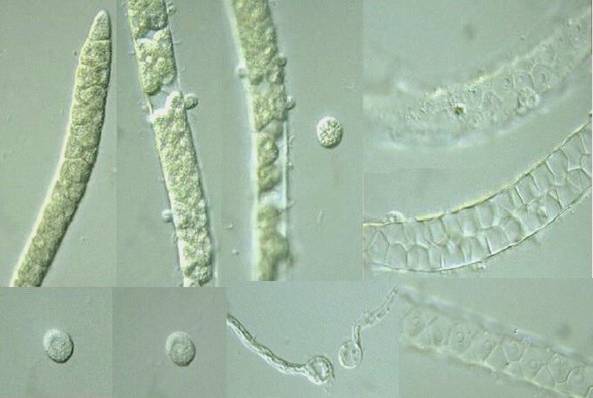
This is the last of the oomycota from my body. I have
found a few of these grooved, coffee bean shaped pieces in the past but
this one was found only a few weeks ago. This zoospore is
Phytophthora infestans,
the villain in the Irish potato famine. This is a motile zoospore powered
by two flagellum. One is a long whiplash flagella and a shorter one is the
shorter tinsel flagella. When these zoospores are threatened they encyst
and can hide out in their encapsulated form for a very long while.
Sometimes they release the flagellum and in other instances they retract the
flagellum and they become encysted as well. The whiplash flagella has a
shape similar to a worm-like structure I often find which I call the motile
strand. The composition of oomycota is cellulose and glucans which store
mycolaminarin and is a form of sugar. If glucans and cellulose are added
to the human body could the results of this addition = excess sugars and
carbohydrates = diabetes? I wonder since I have recently been diagnosed
with it and there is an epidemic of it in this country. I will search for
answers and write a follow up.
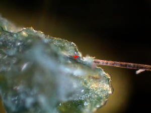
Web photos of phytophthora infestans compared to Morgellons
motile strand.
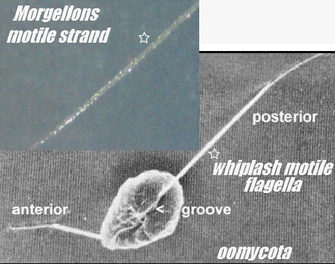
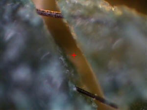
This is what I found in a lesion on my body.
Phytophthora_infestans: Potato blight was one of more than 17 agents
that the United States researched as potential
biological weapons
before the nation suspended its biological weapons program.
France,
Canada,
USA and the
Soviet Union also researched P. infestans as a
biological weapon
in the 1940s and 50s.
Do we really believe that this former candidate as a
biological weapon
was discontinued by the Government? What a
coincidence that it is turning up in the tissue of Morgellons victims.
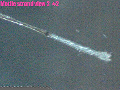
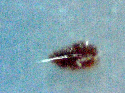
What I do know thus far is that some of the motile (moving)
strands found on my body were tested with Raman Spectroscopy in the labs
at SUNY Stony Brook. The results were surprising to me at the time
since I expected the fibers to be silicon or HDPE (high density polyethylene)
due to previous results of other types of Morgellons samples that had undergone
professional lab analysis.
These strands were made of an indeterminate kind of
polysaccharide (sugar substance- polymer and other unknowns). More
testing was to be done on this material but this was not possible since
the researcher, quite suddenly, was no longer allowed to use their lab. Funny
how any progress on Morgellons research seems to end suddenly as soon as the
research results are posted on the Internet.
I wondered how many sugar/polysaccharide based organisms could
produce motile strands with independent movement. Once again I
could only think of the zoospores of the polysaccharide based oomycota and the
whiplash flagella of pythium insidiosum. There are some chytrids that
produce motile zoospores but not in long stranded fibers. All roads lead
me back to my original conclusion of oomycota.
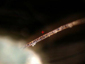
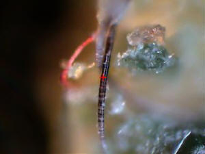
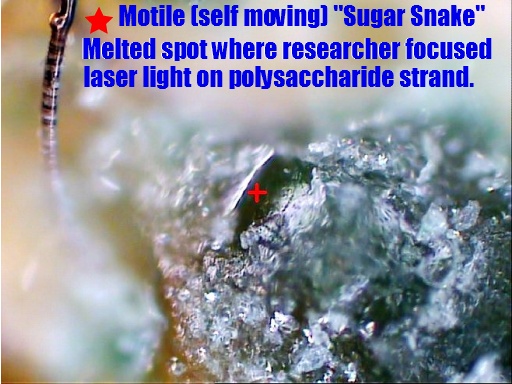
The researcher, Biologist Mark Darrah, nicknamed these Sugar
Snakes. The coloration varies with these strands. They are
sometimes a white crystal-laden translucent coloration and vary to brown or
black as well as striped mixtures of several colors. The high magnification
that was used by Mark Darrah clearly shows a striped banding on the
structures. Mark shined a laser light on one of the strands and it
literally melted. There is a deformity in it's surface at the top left of
the photo below. Here is what he stated to me in about this: �This is a
pimple that formed when the laser light hit it demonstrating heat sensitivity.�
To see more of Mark's work go to:
http://lymebusters.proboards.com/index.cgi?board=research&action=display&thread=10186&page=1
There are 7 pages of his lab photos at this
site.
|
|
|
|
The question I have now is are the materials in these
strands related to the beta glucans in the oomycota? The cellulosic
compounds and Beta-1,3-Glucans in oomycota form a natural polymer in it's
original state. In this bioengineered state what is the purpose of
the material as it is being introduced to our bodies through many
vectors? The answers are rapidly coming into focus.
After further research I have determined that the specific
strains of oomycota that I have found in my body all contain soluble
Beta-1,3-Glucans. This is an important new discovery since out of the
500 species of oomycota only a small percentage contain this
Beta-1,3-Glucans characteristic. All of the oomycota species I
identified as throwbacks have this Beta-1,3-Glucans in their makeup.
What are the odds of
that?
At the original time I did the oomycota research which
started 5 years ago there was no immediate connection I could even imagine
to link my oomycota samples to biotechnology or nanotechnology. I had
not yet found the other nanotechnology items that surfaced in my
body which I have since identified. Read "Living With a
Nightmare"
for more information. It has only
been lately that I revisited the past and decided to see if there was
somehow a nano or bio lab connection with my earlier finds.
These are the types
oomycota I believe I have found
:
Saprolegnia,
Dictyuchus,
Phytophthora.
Peronospora Tabacina
I identified them 5 years ago with no thought in mind to ever
match them to any data. The odds against finding these particular
oomycota specimens and making a meaningful match for them is
astronomical. The article below regarding the surprising
characteristics of Beta-1-3 glucans was a fortuitous find in
my recent research.
"A soluble
Beta-1,3-Glucan Found in Selected Genera of Oomycetes"
From here:
http://mic.sgmjournals.org/cgi/reprint/72/2/393.pdf
�The polysaccharide extracted from each organism was water
soluble and had an infrared spectrum very similar to the glucan of
Phytophthora and Achlya. The infrared spectra of the glucans of Phytophthora, Saprolegnia, Dictyuchus and Pythium are identical."
Further investigation from other sources state that Peronospora Tabacina or blue tobacco mold also contains the
Beta-1,3 Glucans.
http://www.plantphysiol.org/cgi/content/full/117/2/585
Now that I have
established the probable presence of the Beta-1,3-Glucans by way of
oomycota in Morgellons victims, the following article which I
recently found is not a coincidence. The law of averages ran out a
long time ago for this to be coincidental. I believe we are looking
at a huge puzzle piece in the Morgellons mystery.
What could oomycota possibly have to do with biotechnology
or nanotechnology? Please read the following article.
"Beta-1,3-glucan polysaccharides as novel
one-dimensional hosts for DNA/RNA, conjugated polymers and
nanoparticles."
Sakurai K, Uezu K
,
Numata M, Hasegawa
T, Li C
,
Kaneko
K, Shinkai
S
http://www.ncbi.nlm.nih.gov/pubmed/16136229
Department of Chemical Processes and Environments, Faculty
of Environmental Engineering, The University of Kitakyushu, Hibikino, 1-1
Wakamatsu-ku, Kitakyushu, Fukuoka, 808-0135, Japan.
Beta-1,3-glucan polysaccharides have triple-stranded helical
structures whose sense and pitch are comparable to those of
polynucleotides. We recently revealed that the beta-1,3-glucans could
interact with certain polynucleotides to form triple-stranded and helical
macromolecular complexes consisting of two polysaccharide-strands and one
polynucleotide-strand. This unique property of the beta-1,3-glucans
has made it possible to utilize these polysaccharides as potential carriers
for various functional polynucleotides. In particular, cell-uptake
efficiency of the resultant polysaccharide/polynucleotide complexes was
remarkably enhanced when functional groups recognized in a biological
system were introduced as pendent groups. The beta-1,3-glucans can
also interact with various one-dimensional architectures, such as single-walled
carbon nanotubes, to produce unique nanocomposites, in which the
single-walled carbon nanotubes are entrapped within the helical
superstructure of beta-1,3-glucans. Various conductive polymers and gold nanoparticles
are
also entrapped within
the helical superstructure in a similar manner. In addition,
diacetylene monomers entrapped within the helical superstructure can be
photo-polymerized to afford the corresponding poly(diacetylene)-nanofibers
with a uniform diameter.
These
findings indicate that the
beta-1,3-glucans are very attractive and useful materials not
only in biotechnology
but also in nanotechnology.
These unique properties of the beta-1,3-glucans undoubtedly
originate from their inherent, very strong helix-forming character which
has never been observed for other polysaccharides.
(This article was found at pubmed the rest of the article is
only available by subscription)
Note: The number 3 in Beta-1,3 glucans indicates 3
carbon atoms. The next articles is also about the use of sugars
with 3 carbon atoms.
http://www.scientificblogging.com/news_releases/gna_dnas_chemical_cousin_is_a_nanotechnology_building_block
dnas_chemical_cousin_is_a_nanotechnology_building_block
Chemical Cousin of
DNA Provides New Nanotechnology Building Block
�In the case of GNA, the sugar is the only difference with
DNA. The five carbon sugar commonly found in DNA, called deoxyribose,
is substituted by glycerol, which contains just three carbon atoms.�
�The only chemical difference between DNA and a synthetic
cousin, GNA, is in the sugar molecule. GNA uses a three-carbon sugar
called glycerol rather than the five-carbon deoxyribose used in DNA.
The sugar provides the chemical backbone for nucleic acid polymers,
anchoring a phosphate molecule and nitrogenous base (B). Credit: Biodesign
Institute at Arizona State University.�
~~~~~~~~~~~~~~~~~~~~~~~~~~~~~~~~~~~~~~~~~~~~~~~~
On to the worms!
Here are some odd finds I made a few years back. I am
only glad that I have not seen any of these recently.
|
Web
drawing of an earthworm cocoon
|
Cocoon
from inside wrist lesion view 1
|
|
Cocoon
view 2
|
Cocoon
view 3
|
Cocoon
view 4
|
|
Type
2 Cocoon view 1 |
Type
2 Cocoon view 2 |
|
More
clues about earthworm genes 60x
|
Creepy
worm fiber 200x |
It is no surprise to me that people with Morgellons often
find insect-like creatures in their lesions. I believe these
throwbacks can be whatever genetics are part of the brew. It may vary
in different people as to what they may find. It may be a function of
their own genetics as to what manifests. Could these worm genes now
be a part of the polysaccharide �Sugar Snake?� Here are more
mutations to ponder. Which part is the worm and which is the
polysaccharide? Some throwbacks can't make up their minds.
|
Plastic-like
worm |
Polysaccharide Strand with stomach? |
Points to ponder:
Many people with Morgellons disease have noted that they rarely get any bacterial or secondary
infections in the lesions. The dictyostelium are eukaryotic microorganisms that eat bacteria for lunch. (they also produce ammonia).
Normally there would not be enough bacteria on the human body to keep dictyostelium adequately supplied with it's fuel. There have been findings in
Morgellons victims by other researchers of mycoplasma and chlamydya pneumoniae which are both bacteria that are especially difficult to kill on
the human body.
Recent reports have come in from others
with Morgellons disease who state that in clinical testing there is a nearly complete a lack of healthy bacteria in their intestines but there other
harmful bacteria in their bodies.
The need for probiotics and Diflucan or cellulose eating products that contain cellulase and hemicellulase like Candex or NSI Candida yeast Management Capsules becomes necessary. These types of products may be helpful for overgrowth of cellulose combined with the lack of beneficial bacterial flora in the body.
This lack of beneficial bacterial flora would be further evidence of the dictyostelium feeding on the beneficial bacteria in the body. There is anecdotal evidence that that if sugars and carbohydrate consumption are lessened it may help to lessen the negative impact of lesions and symptoms. The pathogen that I believe many have is a mixture of oomycota and the dictyostelium. Just like Candida which has cellulose as a component the oomycota has cell walls of cellulose. The human body has no way to rid itself of cellulose. Feeding the body carbohydrates which will ultimately become sugar overgrowth due to lack of good bacteria in the gut, seems support this cellulose growth. To lessen the cellulose growth, carbohydrates are best kept to a minimum. Since I have had type 2 diabetes this past year and have had to alter my carbohydrate intake, my skin manifestations have lessened. Two other people I know have tried this and have had many of their skin symptoms abate. It is certainly not a cure but it may be helpful to know if skin symptoms are especially aggressive.
Unfortunately the internal nano wiring and and other body symptoms have remained largely unchanged.
Could it be that some symbiotic balance is being kept in the grand scheme of things to create a favorable environment for the dictyostelium, oomycota and other genetic bio-mixtures to live well and prosper? In the bigger picture most things found are serving a purpose and act together. These are not all random things thrown into the mix without thought or purpose. To find the answers, all of the components must be looked at as a whole system.
Along with that we have the presence of Beta-1,3-Glucans from the oomycota varieties which are known for their anti-tumor effects and beneficial effects on the immune system. How nice of them to throw out a health bonus. As I have just mentioned it is also known for it's ability to produce an environment for the development of nano machines and bio-engineering. Take a look at my photo journal and see the many categories of wires and components inhabiting the human body. This is not random.
Consider the implications of the bigger picture and the fact that most people without symptoms have had the same exposure to the same chemtrails and tainted food as Morgellons victims. These people may simply be assimilating the technology without problems or symptoms. Morgellons disease may actually be the lack of ability to assimilate the crap they are dumping on us due to an aberration in our immune systems or our genetics. This aberration was probably unforeseen by those who are doing this to us. As such it is an unwelcome circumstance that is being ignored, ridiculed and constantly monitored by professionals. Reality is not always apparent until it is in hindsight. Further, all "people" we meet are not necessarily what they appear to be. If all is as I suppose it to be considering what I see coming out of my own body on a daily basis, there would be a small army in place to contain the Morgellons information from the public.
Morgellons is not a one-component disease but a system of synergistic pathogens with a purpose.
As an update, I want the public to understand that there is no valid funded research being done by our government. A handful of people are trying to make progress with their own time and money but it seems that there is a brick wall placed in front of us at every turn. I believe the term for this is that we are being "contained". A number of medical professionals have found that involvement with the Morgellons issue has led to government shakedowns of their records in attempts to find any reason to close them down. Other researchers have been threatened and more. There is a dark underbelly of evil that is calling the shots regarding this man-made disease. It is time to wake up and discern the truth.
Let's spread the truth and the love - they just hate that.
"Greema
10-13-2009
Source: http://www.morgellonsexposed.com/Throwback.htm