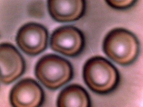
One of the more normal appearing regions of red blood cells from a nine-year old child.
Magnification approx. 7000x
2. The vast majority of all blood samples observed (if not all) are showing various degrees of anomalous form. The degree of damage to cellular integrity and form appears to correspond directly to the number of anomalous structures that are found within any individual sample.
3. The anomalies in a variety of blood samples have transcended the age factor, and they have now also been observed in the same fashion and form within the blood of a nine year old child.
In brief, the method of assessment is as follows:
1. The organism involved must measure on the order of sub-micron, generally on the order of 0.5 to 0.7 microns.
2. The organism is an intra-cellular organism, and can exist within human red blood cells (erythrocytes).
3. The organism is likely to be associated with respiratory conditions and is circulated by aerosol means.
4. The organism is spherical to oblate, but can can exist in more than one form (pleomorphic).
5. Gram stain procedures applied to the blood samples should produce a gram-negative result.
6. The same form, at least by inspection and measurement, should in general be present in diverse Morgellon physical samples, the airborne fibers under long term investigation(EPA refused) and the blood of Morgellon individuals. The general population may also demonstrate a marked presence of the pathogen.
7. The illnesses that are associated with the Chlamydiae genus should correlate with increasing and pervasive ailments and diseases.
8. Recorded observations of the pathogen must match the accumulated observations of this researcher.
Considerable attention has been given to the consideration of alternative explanations, such as blood platelet imbalances, conventional bacterial and fungal forms; contradictions to one or more of the above conditions occur during those analyses. Although all pathogens remain under consideration a focus on Chlamydia-like groups is in place. The Chlamydiae-like group(with a special interest in Chlamydia pneumoniae) satisfies the above conditions to the degree that is understood. Again, this assessment is not intended to be final, but it is offered as the best assessment by this resarcher to date. It is not unexpected that modification of a Chlamydia group could take place(e.g., mycoplasma combinations). In addition, the relationships to the second structural form(sub-micron filament network and bounding filaments) will need to be provided.
One of the more normal appearing regions of red blood
cells from a nine-year old child.
Magnification approx. 7000x
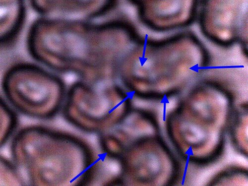
Anomalous region of blood cells of nine
year old child prior to the gram stain process.
Cellular
integrity damage is apparent. If cellular variation is extensive in the
sample,
sub-micron structures are often visible(blue arrows) without
any staining process involved.
Degree
of cellular damage appears to correspond directly to the number of chlamydia-like structures
within,
in contact with or adjacent to the blood cells.
Numerous
"double"-erthyrocte forms observed in this sample.
Magnification approx.
7000x..
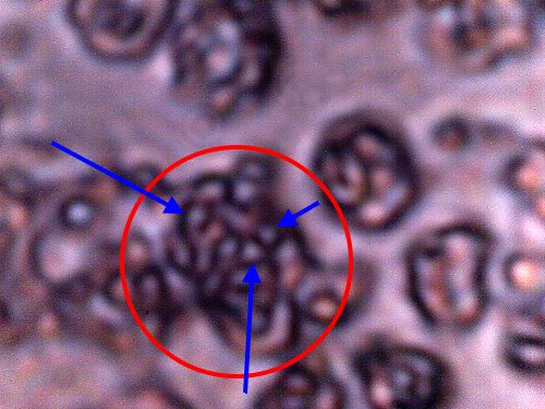
Red blood cell of nine year old child after the gram
stain process;
visible examination of slide appears to show the gram
negative process as highly dominant.
Area within circle
encloses erthyrocte(red blood cell) boundary prior to stain
process.
Blood cell structure is damaged during the stain
process(expected).
Certain bacterial
types or bacterial-like types(blue arrows) remain and
are emphasized after the stain process.
The
intra-cellular (chylamidia-like) structures measure approx. 0.5 -
1.0 microns in diameter.
Due to morphological
variation across various samples observed,
crossovers with mycoplasma forms
(also pleopmorphic) remain under strong consideration.
Magnification approx. 7000x.